Kategórie
- Novinky
- MŠ a predškolské vyučovanie
- Prvý stupeň ZŠ
- Druhý stupeň ZŠ
- Slovenská jazyk a literatúra
- Cudzie jazyky
- Matematika
- Informatika
- Fyzika
- Chémia
- Biológia
- Dejepis
- Geografia
- Občianska náuka
- Hudobná výchova
- Výtvarná výchova
- Výchova umením
- Etická výchova, náboženská výchova
- Technická výchova
- Environmentálna výchova
- Telesná a športová výchova
- Dopravná výchova
- Ochrana života a zdravia
- SŠ a odborné vyučovanie
- Odborné učebne a knižnice
- Výučbové softvéry
- Interaktívna technika
- Školské tabule
- Školský nábytok
- SKLADOVKY
TOP PRODUKTY
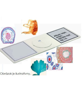
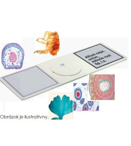
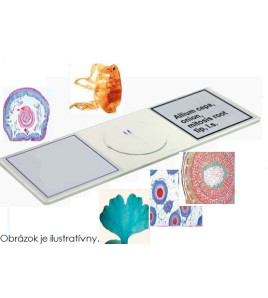
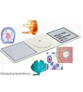
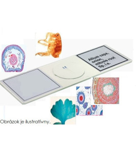
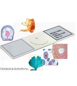
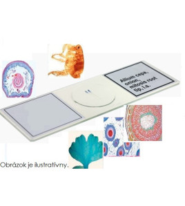
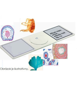
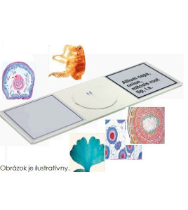
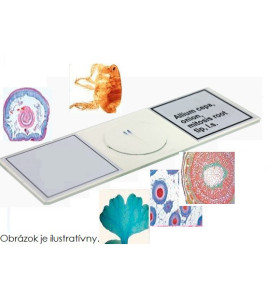
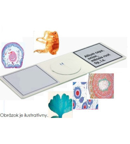
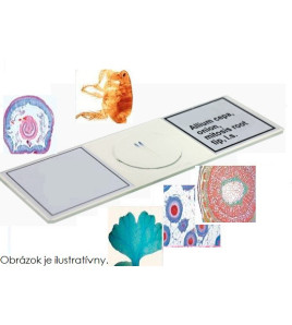
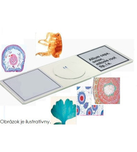
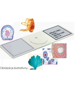
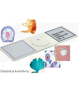
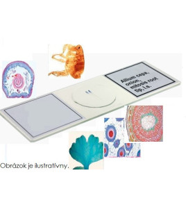
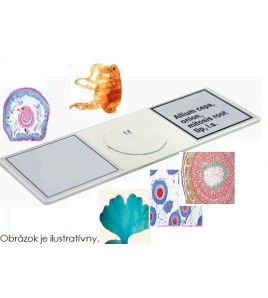
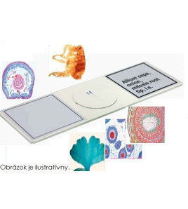
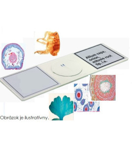
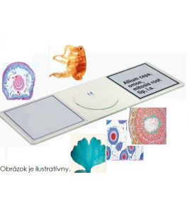
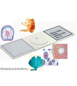
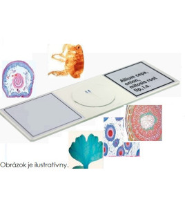
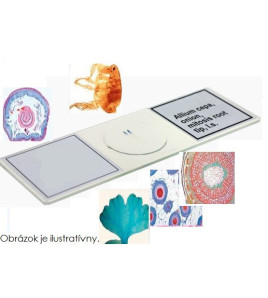
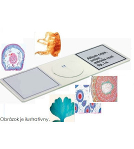
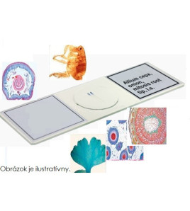
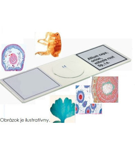
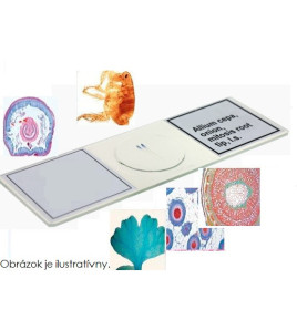
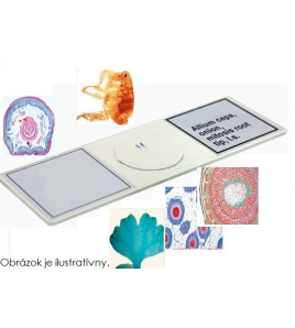
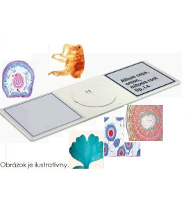
Mikroskopické preparáty - Školská sada B, 50 preparátov
12000576D
590,40 € s DPH
s DPH
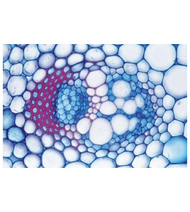
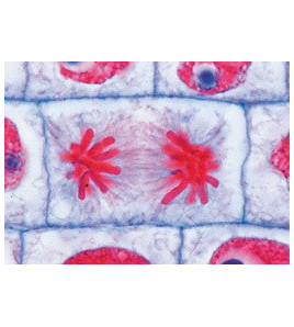
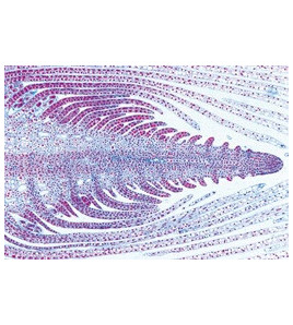
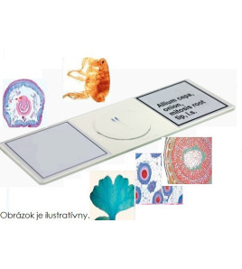
Mikroskopické preparáty - Školská sada B, 50 preparátov (zoológia a parazitológia, histológia človeka a cicavcov, baktérie a rastliny).
Dostupnosť: Na sklade u dodávateľa, dodanie do 30 dní
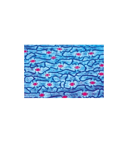
Mikroskopické preparáty - Školská sada B, 50 preparátov (zoológia a parazitológia, histológia človeka a cicavcov, baktérie a rastliny). Zoology and Parasitology: 1(d) Paramaecium, nuclei stained 2(c) Euglena, a common flagellate with eyespot 3(c) Sycon, a marine sponge, t.s. of body 4(e) Dicrocoelium lanceolatum, sheep liver fluke, w.m. 5(c) Taenia saginata, tapeworm, proglottids of various ages t.s. 6(d) Trichinella spiralis, l.s. of skeletal muscle showing encysted larvae 7(d) Ascaris, roundworm, t.s. of female in region of gonads 8(b) Araneus, spider, leg with comb w.m. 9(d) Araneus, spider, spinneret w.m.10(d) Apis mellifica, honey bee, mouth parts of worker w.m. 11(b) Apis mellifica, hind leg of worker with pollen basket w.m. 12(e) Periplaneta, cockroach, chewing mouth parts w.m. 13(b) Trachea from insect w.m. 14(b) Spiracle from insect w.m. 15(d) Apis mellifica, sting and poison sac w.m. 16(b) Pieris, butterfly, portion of wing with scales w.m.17(d) Asterias rubens, starfish, arm (ray) t.s. showing tube feet, digestive gland, ampullae.Histology of Man and Mammals: 18(e) Fibrous connective tissue of mammal 19(c) Hyaline cartilage of mammal, t.s. 20(e) Adipose tissue, stained for fat 21(d) Smooth (involuntary) muscle l.s. and t.s. 22(e) Medullated nerve fibres, teased preparation of osmic acid fixed material showing Ranvier’s nodes 23(c) Frog blood smear, showing nucleated red corpuscles 24(d) Artery and vein of mammal, t.s. 25(d) Liver of pig, t.s. showing well developed connective tissue 26(c) Small intestine of cat, t.s. showing mucous membrane 27(c) Lung of cat, t.s. showing alveoli, bronchial tubes.Cryptogams: 28(c) Oscillatoria, a common blue green filamentous alga 29(e) Spirogyra in scalariform conjugation, formation of zygotes 30(c) Psalliota, mushroom, t.s. of pileus with basidia and spores 31(c) Morchella, morel, t.s. of fruiting body with asci and spores 32(d) Marchantia, liverwort, antheridial branch with antheridia l.s. 33(d) Marchantia, archegonial branch with archegonia l.s. 34(d) Pteridium, braken fern, rhizome with vascular bundles t.s. 35(d) Aspidium, t.s. of leaf with sori showing sporangia and spores.Phanerogams: 36(e) Elodea, waterweed, stem apex l.s. showing meristematic tissue and leaf origin 37(d) Dahlia, t.s. of tuber with inuline crystals 38(b) Allium cepa, onion, w.m. of dry scale showing calcium oxalate crystals 39(d) Pyrus, pear, t.s. of fruit showing stone cells 40(c) Zea mays, corn, typical monocot root t.s. 41(c) Tilia, lime, woody dicot root t.s. 42(c) Solanum tuberosum, potato, t.s. of tuber with starch and cork cells 43(c) Aristolochia, birthwort, one year stem t.s. 44(c) Aristolochia, older stem t.s. shows secondary rowth 45(d) Cucurbita, pumpkin, l.s. of stem with sieve tubes, annular and reticulate vessels, sclerenchyme fibres 46(d) Root tip and root hairs 47(c) Tulipa, tulip, epidermis of leaf with stomata and guard cells w.m., surface view 48(c) Iris, typical monocot isobilateral leaf, t.s. 49(c) Sambucus, elderberry, stem showing lenticells and cork cambium, t.s. 50(e) Triticum, wheat, grain (seed) sagittal l.s. with embryo and endosperm.


